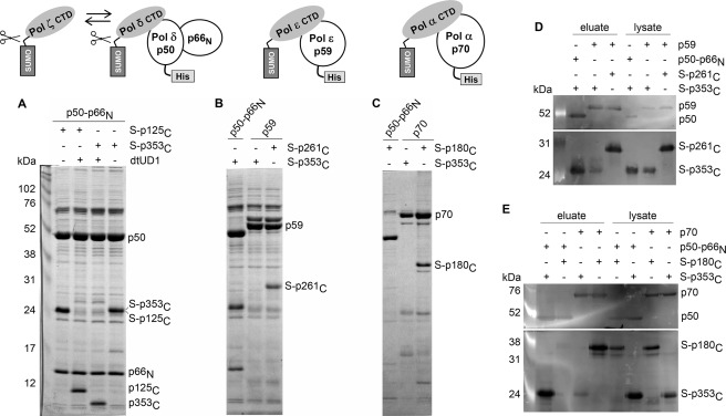
FIGURE 3.
Analysis of interaction between B-subunits and CTDs of human DNA polymerases. Samples loaded on and eluted from Ni-IDA were subjected to 12% SDS-PAGE, followed by detection with Coomassie Blue staining (A–C) or by Western blotting (D and E). A. The Pol δ B-subunit binds to the CTDs of Pol δ and Pol ζ. The SUMO (S) tag was cleaved off by dtUD1 prior to binding to the resin. B and D, the Pol ϵ B-subunit binds to the CTD of Pol ϵ and not to the CTD of Pol ζ. C and E, the Pol α B-subunit binds to the CTD of Pol α and not to the CTD of Pol ζ. The Pol δ B-subunit does not bind to the CTD of Pol α. Left lanes in A, D, and E, ECL Plex fluorescent rainbow markers (GE Healthcare). DNA Pol subcomplexes are schematically shown above the A–C. All B-subunits have N-terminal His6 tags.

