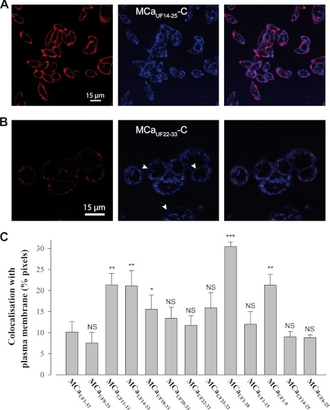
FIGURE 4.
Membrane staining is diffuse whereas intracellular staining is punctuated. A, lower magnification image of CHO cells stained with 3 μm MCaUF14–25-C-Cy5 that illustrates a predominant subplasma membrane rim-like distribution. B, diffuse membrane staining of CHO cells by MCaUF22–33-C-Cy5. White arrows indicate domains of the plasma membrane where the diffuse staining of the peptide-cargo complex is the most evident. C, extent of colocalization of the Cy5-labeled peptides with the rhodamine-labeled plasma membrane. NS, nonsignificant; *, ≤0.1; **, ≤0.05; and ***, ≤0.001.

