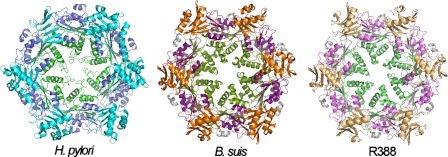
FIGURE 7.
Hexameric structures of VirB11 proteins. The hexameric structures of H. pylori HP0525 (1nlz.pdb), B. suis VirB11 (2gza.pdb), and R388 TrwD (see “Experimental Procedures”) are shown. Each monomer consists of an NTD (cyan, orange, and wheat, respectively) and a CTD (blue, magenta, and pink, respectively), connected by a flexible linker (gray). The region at the C-terminal end identified by papain proteolysis in TrwD (green) and the equivalent positions in B. suis VirB11 (olive) and HP0525 (lime) is facing the interior of the hexameric rings.

