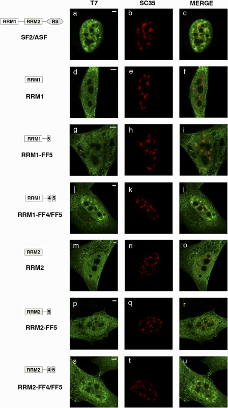
FIGURE 3.
FF4/FF5 directs SRSF1 domain-deletion mutants to nuclear speckles. Cells were transfected with the indicated plasmids and dually labeled with antibodies directed against the expressed SRSF1 protein (left column, green) and SC35 (center column, red). The merged images are also shown (right column). In all cases, colocalization of expressed proteins with the endogenous marker was assessed by confocal imaging. A diagrammatic representation of the T7-tagged SRSF1 mutants used is shown at the left of the figure. The structure of the SRSF1 domain-deletion mutants was described previously (51). Scale bars = 3 μm.

