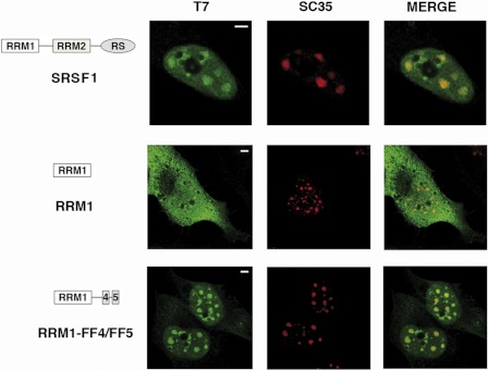
FIGURE 4.
Colocalization of the FF4/FF5-containing RRM1 protein with nuclear speckles is not perturbed following inhibition of transcription. HeLa cells were transfected with the indicated expression plasmids and treated with 25 μg/ml of α-amanitin for 6 h at 37 °C and then processed for immunofluorescence analysis. Dual-labeling of cells with antibodies directed against SRSF1 (T7, green) and with the SC35 antibody (red) was performed. Individual staining and merge images of the cell stained with the indicated antibodies are shown. A diagrammatic representation of the T7-tagged SRSF1 mutants used is shown at the left of the figure. Scale bars = 3 μm.

