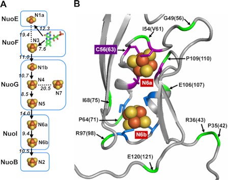
FIGURE 1.
Arrangement of Fe/S clusters in E. coli NDH-1. A, schematic drawing of all Fe/S centers and cofactor FMN displaying their locations in the subunits. Edge to edge distances are given. Postulated flow of electrons through the main pathway is indicated with arrows. B, N6a and N6b clusters and their surroundings in the Nqo9 subunit (E. coli NuoI homolog) from hydrophilic domain of T. thermophilus complex I (Protein Data Bank code 3IAS). The coordinating cysteine residues for the clusters N6a and N6b are highlighted in purple and blue, respectively. The locations of some of the selected residues used for mutations in the vicinity of the two clusters are shown in green and marked by an arrow with their T. thermophilus numbering. In addition, E. coli numbering is displayed in the parentheses. It should be noted that Ile-54 in T. thermophilus is replaced by Val-61 in E. coli.

