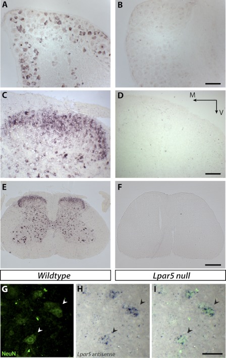
FIGURE 2.
In situ hybridization and immunolabeling of tissues from wild type and null mutant mice with Lpar5 digoxigenin-labeled antisense probes and NeuN antibody. A and B, sections of DRG from wild type (A) and Lpar5 null mice (B). C and D, sections of spinal cord dorsal horn from wild type (C) and Lpar5 null mice (D) show Lpar5 expression in the dorsal horn area. Scale bar, 100 μm. M, medial; V, ventral. E and F, low magnification images of whole spinal cord sections from wild type (E) and Lpar5 null mice (F). Note that both dorsal horn and ventral horn neurons are labeled. Scale bar, 400 μm. G–I, anti-NeuN antibody immunostaining (G) and in situ hybridization against Lpar5 (H) confirmed Lpar5 expression in double labeled neurons (I). The arrowheads indicate the same cells from G–I. Scale bar, 50 μm.

