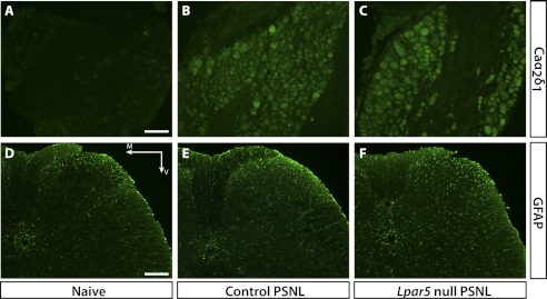
FIGURE 4.
Immunohistochemistry showing Caα2δ1 immunoreactivity in DRG as well as GFAP immunoreactivity in the L5 spinal cord region 6 days after PSNL. A–C, Caα2δ1 is significantly up-regulated in DRG after PSNL; however, no significant difference was observed between heterozygous control and null mutant mice. Scale bar, 100 μm. D–F, GFAP was significantly increased in PSNL mice, indicating astrocyte activation; however, Lpar5 null mutant mice showed similar, if not increased, GFAP immunostaining compared with the heterozygous control. Scale bar, 200 μm. M, medial; V, ventral.

