FIGURE 7.
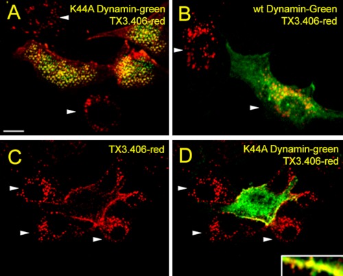
Role of dynamin in ET-1 induced internalization of TX3.406 in cultured endothelial cells. Images from confocal fluorescence microscopy of RLMVEC already transfected to express either K44A dynamin2-eGFP (green) (A, C, and D) or wild type (wt) dynamin2-eGFP (green) (B). The cells were incubated with TX3.406 for 1 h at 4 °C and washed before 5 min of stimulation with ET-1 at 37 °C and then fixation, permeabilization, and incubation with secondary fluorescent reporter antibody to mouse IgG (red). The arrowheads indicated intracellular perinuclear staining. Bar, 10 μm.

