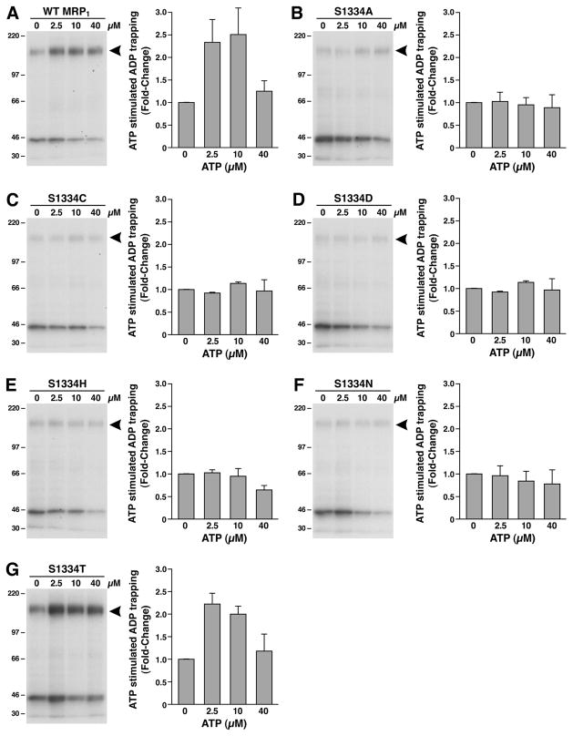
Figure 6.
Substitution of S1334 in the Walker A motif of NBD2 with an amino acid residue that eliminates the hydroxyl group abolished the ATP-enhanced vanadate-dependent ADP trapping at the mutated NBD2. The photolabeling was carried out in 10 μL of solution containing 10 mM MgCl2, 800 μM vanadate, 5 μM [α-32P]-8-N3ADP, varying concentrations of ATP as indicated on top of the gel, and 10 μg of MRP1-containing membrane vesicles. The reaction mixture was incubated at 37 °C for 10 min, brought back to ice, washed with 500 μL of ice-cold Tris–EGTA buffer (0.1 mM EGTA and 40 mM Tris-HCl, pH 7.5), and UV-irradiated (λ = 254 nm) on ice for 2 min. The labeled proteins were separated on a 7% polyacrylamide gel and electroblotted to a nitrocellulose membrane. The arrowhead on the right of the gel indicates the photolabeled MRP1 protein. The counts in each MRP1 band were measured by Instant Imager, and the amount of labeling in the absence of ATP was considered as 1 (n = 3).

