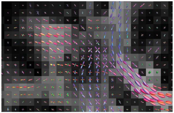
FIG. 14.
Fiber orientation distributions for each voxel, for a coronal section showing the lateral projections of the corpus callosum (left-right: red lobes) crossing through the fibers of the corona radiata (inferior–superior: blue lobes) and of the superior longitudinal fasciculus (anterior–posterior: green lobes). Results produced from data obtained from a healthy volunteer, consisting of 60 DW directions acquired at b = 3000 s/mm2 (9 min scan time), analyzed using constrained spherical deconvolution (99).

