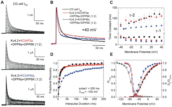
Figure 7. Reconstitution of native ISA channel from CG cells by heterologous expression in oocytes.
(A) Outward transient currents elicited from CG cells and oocytes expressing Kv4.2, a mixture of DPP6a and DPP6K at 1∶2 ratio, and either KChIP3a or KChIP4bL. From a holding potential of −100 mV, either a 200-ms (CG cells) or 1-sec (oocytes) step depolarizations were made from −100 mV to +40 mV at 10 mV increments. (B) Overlapped normalized current traces at +40 mV from the indicated channels. (C) Time constants of inactivation at indicated membrane potentials for ISA from CG cells, Kv4.2+KChIP3a+DPP6a+DPP6K (1∶2), and Kv4.2+KChIP4bL+DPP6a+DPP6K (1∶2). (D) Recovery from inactivation at −100 mV, measured using the two-pulse protocol. (E) Normalized peak conductance-voltage relations (Gp/Gp,max) and steady-state inactivation curves (I/Imax) for ISA from CG cells and reconstituted channel complexes.

