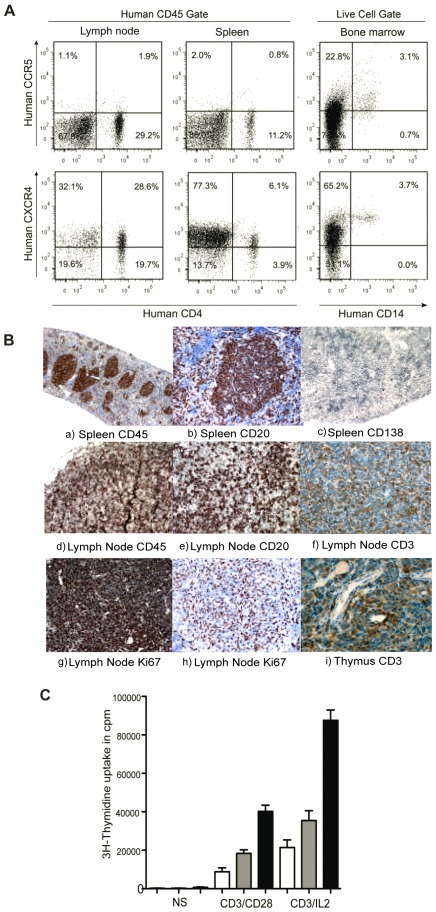
Figure 3. Coreceptor expression and lymphoid organ formation in humanized mice.
(A) Representative FACS profile of human CCR5+CD4+ and CXCR4+CD4+ cells in spleen and lymph nodes, gate was set of human CD45 cells. Expression of CCR5 and CXCR4 were checked on CD14+ cells, gate was set on live human cell population. (B) Histology of lymphoid organs in CD34+ engrafted NSG mice. The lymphoid follicles mainly contained hCD45 cells. Spleen sections were stained with (a) anti-hCD45, (b) anti-hCD20 and (c) anti-hCD138. Lymph node sections were also stained with (d) anti-hCD45, (e) anti-hCD20 (f) anti-hCD3 (g,h) anti-hKi67 antibodies. Thymus section was stained with anti-CD3 antibody. (C) Proliferative responses of T cells were measured in nonstimulated cells, after stimulation with immobilized anti-CD3/anti-CD28 and anti-CD3+IL2 in spleen (white bars), lymph nodes (gray bars) and human PBMCs (black bars).

