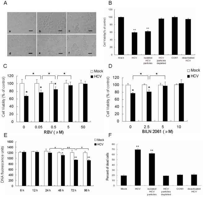
Figure 2. HCV played a direct role in the reduction of cell viability and induction of cell death of beta cells.
(A, B) MIN6 cells were mock-infected (a) or infected with 1.0 MOI of the supernatant of HCV-infected Huh7.5.1 (b), ultracentrifugation-purified HCV particles (1.0 MOI) (c), the supernatant of HCV-infected Huh7.5.1 after ultracentrifugation (d), CON1 cell culture medium (e), or the supernatant of HCV-infected Huh7.5.1 after UV radiation treatment (f) at 96 hpi. Light microscopy images (scale bar, 10 µm) (A) were obtained and cell viability (B) of MIN6 cells was determined by MTT assay. (C, D) MIN6 cells were mock-infected or infected with HCV particles (1.0 MOI) with addition of indicated dose of RBV (C) or BILN 2061 (D) at 96 hpi, cell viability was assessed by MTT assay. (E) MIN6 cells were infected with HCV as in Figure 1B. At different time-points, cell number was determined by DNA content staining with the fluorescent dye PI. (F) MIN6 cells were treated as in (A). Cell death induced by the indicated treatment was measured by trypan blue exclusion. (A–F) All activity measurements were done in triplicates. Data represent means + SD of three independent experiments (n = 9). *P<0.05, **P<0.01, compared with respective controls.

