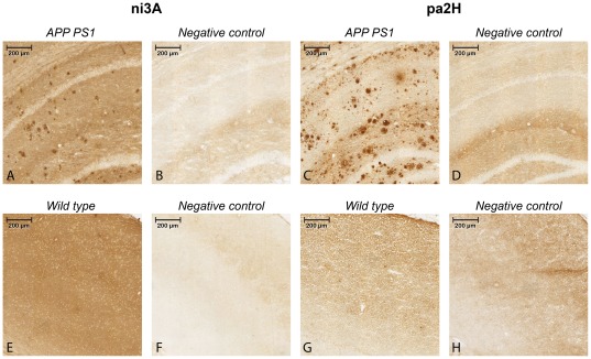
Figure 1. Immunostaining on murine APP/PS1 sections using ni3A and pa2H.
The upper panels (A–D) show 10× magnifications of the resulting staining with cryosections of aged APP/PS1 mouse brain tissue including negative controls, while the lower panels (E–H) show similar staining performed with wildtype littermates.

