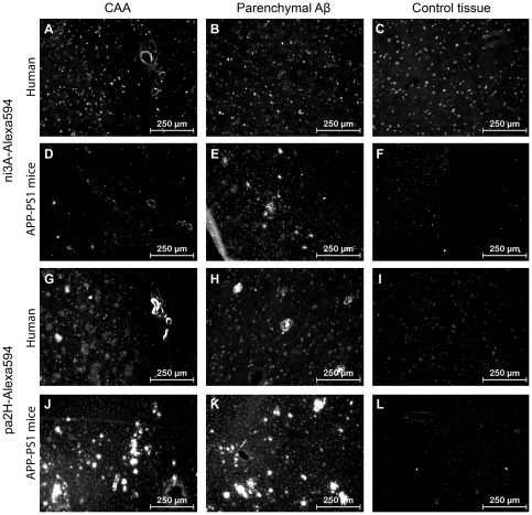
Figure 5. Immunofluorescence with VHH-Alexa594.
Shown are the results of immunofluorescence staining with ni3A- and pa2H-Alexa594 on cryosections of APP/PS1 murine and human AD/CAA brain tissue, including wildtype or healthy controls. Both VHH stain positive for CAA in all sections (A, D, G, J). Only ni3A-Alexa594 stained negative for human parenchymal Aβ (B), while pa2H stained positive for several types of parenchymal Aβ deposits (G, H, J, K) in both humans and mice. In either human of murine control tissue no such staining patterns were observed.(C, F, I, L)

