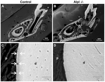
Figure 3. Lack of acellular cementum on Alpl −/− molar root surfaces.
Backcattered SEM was employed to explore the cervical root surface (white boxes) in (A) Alpl +/+ control and (B) Alpl −/− first molars. At higher magnification, the acellular cementum layer (white arrows) in the (C) control molar can be distinguished by contrast differences due to slightly lower mineralization than underlying dentin (d). (D) No acellular cementum layer was apparent in the cervical region of the Alpl −/− molar. Abbreviations: d = dentin; c = acellular cementum; p = periodontal ligament.

