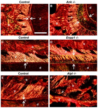
Figure 6. Progressive mineralization of extrinsic collagen fibers in Ank and Enpp1 −/− cervical cementum.
Picrosirius red staining with polarized light microscopy was used to visualize birefringent collagen fibers of periodontal tissues in mandibular first molar roots. Histological sections of 60 dpn (A) control Ank; Enpp1 +/+ cut in a horizontal plane and (C) coronal plane revealed high density of embedded extrinsic fibers in the acellular cementum, where the high degree of birefringence (intense coloration) makes visible the organization and orientation of the major PDL collagen fibers. Observation of (B) Ank −/− and (D) Enpp1 −/− expanded cervical cementum (yellow dotted outline, flanked by white arrows) in the same orientations revealed a similar high density of embedded fibers, continuous from PDL through the cementum. (E) Control Alpl +/+ molars at 21 dpn cut in a coronal plane show an organized and attached PDL, while conversely, (F) Alpl −/− molars exhibited tearing at the root-PDL interface (#), osteoid invasion of the PDL space, and poorly organized and sparsely embedded collagen fibers at the cervical root. Abbreviations: d = dentin; c = acellular cementum; p = periodontal ligament; b = bone. Scale bar = 100 µm.

