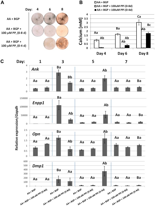
Figure 13. Timing of pyrophosphate removal determines cementoblast mineralization and coordinated gene expression in vitro.
(A) By von Kossa staining, OCCM.30 cells cultured with 5 mM BGP produced mineral nodules by days 4, 6, and 8, while inclusion of 100 µM PPi inhibited mineralization for the entire experiment. When PPi was removed after 4 days, OCCM.30 cells began mineralizing the matrix by days 6 and 8. (B) Quantitative calcium assay performed on days 4, 6, and 8 confirmed visual mineral nodule staining by von Kossa. (C) Mineralizing cultures (AA + BGP) increased expression of Ank, Enpp1, Opn, and Dmp1 at day 3, concurrent with mineralization. Inclusion of 100 µM PPi significantly depressed expression of Ank, Enpp1, and Opn on day 3 compared to mineralizing cultures. Removal of PPi on day 4 led to increased Ank, Enpp1, Opn, and Dmp1 on day 5, coincident with mineralization. Graphs in (B) and (C) show mean +/− SD for n = 3 samples. Lowercase letters indicate treatment comparison at each time point, where different letters indicate a statistically significant intergroup difference. Uppercase letters indicate comparisons over time in the same treatment group, where different letters indicate a statistically significant intragroup difference. Values sharing the same uppercase or lowercase letter in were not significantly different. Means were compared by ANOVA (p<0.05) followed by the Tukey test for direct pair-wise comparisons.

