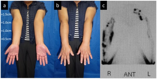
Figure 1. Case 3.
(a, b) Edematous condition before LVA. Compared to the healthy side (left), the circumferences were increased by 1.5, 1.0, 1.0, and 0.5 cm for the upper arm, elbow, forearm, and wrist, respectively. (c) Findings from lymphoscintigraphy. Reflux in the skin (dermal backflow) was observed in the medial and lateral upper arm and lateral forearm in the right upper limb. A normal lymph vessel distribution was apparent in the left upper limb.

