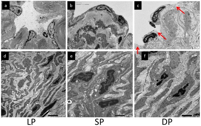
Figure 3. Case 3.
Collecting lymph vascular endothelial cells in (a) linear pattern (LP), (b) splash pattern (SP), and (c) diffuse pattern (DP) areas. Regions in which endothelial cells were detached are indicated with red arrows. (d) ‘Contractile’ smooth muscle cells in (d) LP, (e) SP, and (f) DP areas. The space between smooth muscle cells was widened due to overgrowth of collagen fibers.

