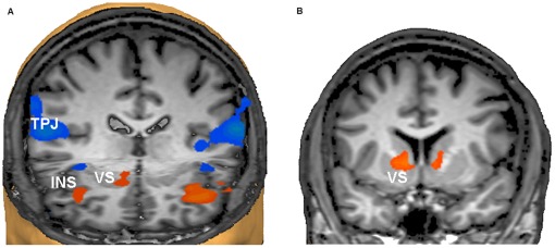
Figure 5. Network activation and deactivation during neurofeedback.
a) Activation of the insular cortex (INS) bilaterally and the right ventral striatum (VS) supported the neurofeedback task, whereas the temporoparietal junctions (TPJ) of both hemispheres were deactivated. The TPJ is recognised as part of the brain’s “default mode network” that is deactivated during effortful tasks. For a full documentation of the activated and deactivated networks see Table 3. View from the front and above. The right side of the brain is on the observer’s left (Talairach coordinates of virtual cuts: y = 25, z = −2). b) Successive training sessions produced further increases of activation during upregulation periods in the VS bilaterally (coronal view at y = 7, the right side of the brain is on the observer’s left).

