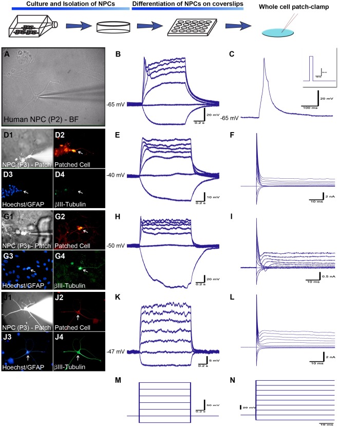
Figure 2. NPCs differentiated on PDL/Laminin-coated coverslips give rise to cells showing neurophysiological properties.
(A-C) Bright field image of a differentiated NPC during patch-clamp recordings and the electrophysiological responses to multiple and single current steps respectively (left to right). This cell had a RMP of −65 mV and fired an immature action potential (AP) in response to depolarizing current. The insert in (C) was the stimulus protocol. (D–F) AhNPC differentiated for 5 days. This cell did not express βIII-tubulin or GFAP, but had clear neurite processes (D1–4). It exhibited a small depolarization and a small inward current in response to depolarizing stimuli (E–F). (G–L) AhNPCs recorded after 3–4 weeks of differentiation. (G–I) Recording from a βIII-tubulin positive, GFAP-negative cell (D3–D4) that fired an immature APs (H), and exhibited small, fast-inactivating inward and slow-inactivating outward currents (I). (J1–4) An ‘asteron’ cell expressing both GFAP and βIII-tubulin. These cells failed to elicit any active voltage or current responses to depolarizing stimuli (K-L). (M-N) The stimulus protocols for current clamp (M) and voltage clamp (N). All traces are representative recordings from 5 different biopsy cases that were conducted from either passage 2 or 3 cells.

