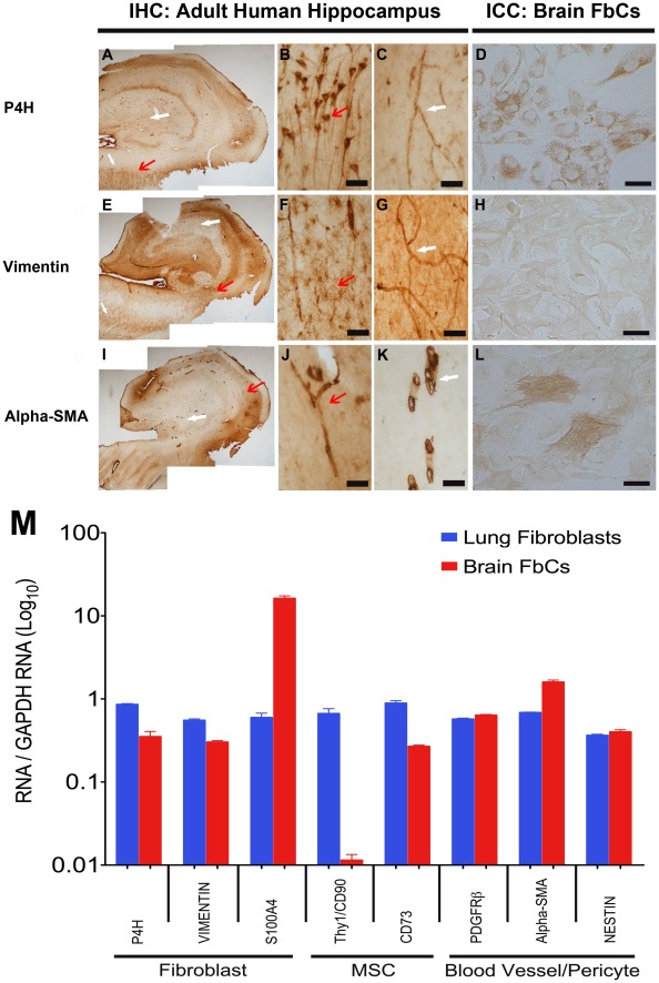
Figure 5. The brain-derived FbCs isolated from biopsy specimens are likely to be of neurovasculature origin.
DAB immunohistochemistry of adult human hippocampus sections and immunocytochemistry of the isolated cells showing positive staining for prolyl-4-hyroxylase (P4H; A–D), Vimentin (E–H) and alpha-smooth muscle actin (Alpha-SMA; I-L). Higher magnification images show P4H to be localized in the pyramidal cells of the CA1 region (A, B; red arrow) and in the blood vessels throughout the section (A, C; white arrow). Vimentin was largely localized to the astrocytes in the white matter (including stratum radiatum; E-F; red arrow), and again, the blood vessels (E-G; white arrow). Alpha-SMA staining was only visible in the blood vessels (I-K; all arrows). Scale: (B-C, F-G, J-K) = 60 µm, (D, H, L) = 100 µm. (M) Comparative analysis of gene expression using q-RT-PCR for FbCs and fibroblast cells isolated and grown from the adult human brain and lung respectively. The level of expression was measured using a standard curve and normalized to a house-keeping gene, GAPDH. From the graph, it is evident that our brain-derived FbCs have a fibroblast-like gene expression profile that appears to be more vasculature cell-like than mescenchymal stem cell (MSC)-like.

