
Figure 1. Dorsal view of the cerebral hemisphere of C57BL/6J wild-type, MMP-9 KO, MMP-2 KO and MMP-2/9 dKO mice after microvascular injection with India ink-stained latex (n = 4 for each group).
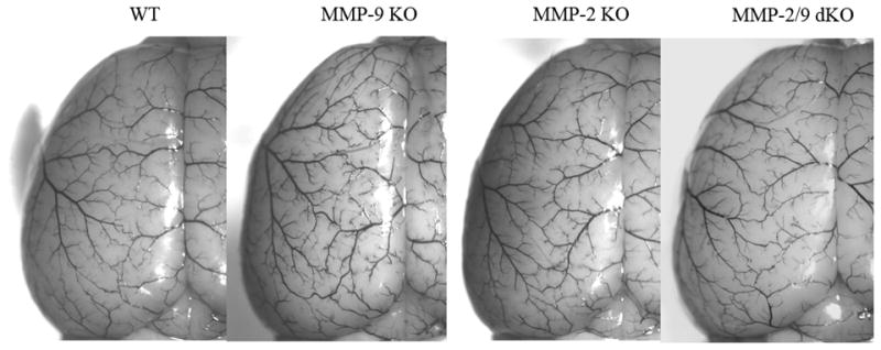
There is a similar distribution of dorsal vessels and boundary zones for the anterior and middle cerebral arteries in all groups.