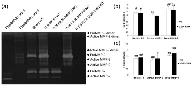
Figure 2. Gel zymogram and quantification of band density in the brains of adult mice after focal MCA ischemia (n = 4).
I, ischemia; R, reperfusion; dKO, double knockout. #p < 0.05, ##p < 0.01 compared with the sham group. a) representative images. b) and c) quantification of MMP-2 and -9 levels. MCA ischemia for 1.5 h followed by reperfusion for 6.5 h leads to increased proMMP-2 and proMMP-9 levels and increased MMP-2 and -9 activation (a, b and c). MMP-9 KO did not significantly increase MMP-2 levels (b), and MMP-2 KO did not significantly increase MMP-9 levels (c) when compared with the WT group after focal MCA ischemia.

