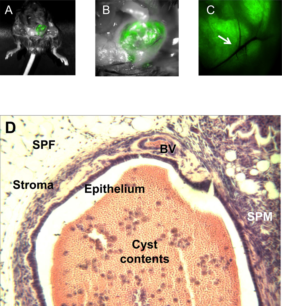
Figure 1.
Endometriotic lesions in recipient mice, 14 d after inoculation. A. GFP+ lesions were visible in situ using interference filter eyeglasses (excitation wavelength, 488 nm; emission wavelength, 502 nm). B. Higher power (5.6x) view shows large cystic and two small satellite GFP+ lesions invading anterior parietal peritoneum and preperitoneal fat in the left lower pelvis. C. High magnification (12x) view of GFP+ lesion shows vascular arcade of established GFP+ implant with arrow indicating vessel branchpoint. Pictures were taken with a Hamamatsu Multiplier CCD Camera (Bridgewater, NJ). D. H&E histology of murine endometriotic lesions was nearly identical to that observed in the human counterpart. This example of a cystic lesion was lined by dense epithelium and surrounded by endometrial stroma. The lesion invaded subperitoneal fat (SPF) and subperitoneal muscle (SPM). The cyst wall contained blood vessels (BV) and the cyst contents included erythrocytes (pink) and leukocytes (purple). Magnification = 40x.

