Figure 4.
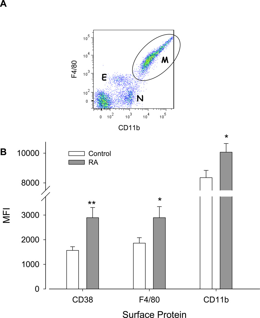
A. Representative dot plot showing CD11b and F4/80 fluorescent intensity of peritoneal cells from a control mouse 14 days after induction of endometriosis. Subpopulations of viable eosinophils (E, CD11bmediumF4/80medium), neutrophils (N, CD11bmediumF4/80negative), and macrophages (M, CD11bhighF4/80high) are indicated. Dot blot analysis of RA-treated mice showed similar population profiles. B. Cells gated on the macrophage (M) population were analyzed for CD11b, F4/80, and CD38 expression. Upregulation by RA of the macrophage markers (CD11b and F4/80) and the differentiation/activation marker CD38 was observed. Values represent the MFI ± SEM of the surface proteins as indicated in control (n=22) and RA-treated (n=22) mice. *, p< 0.03; **, p<0.002.

