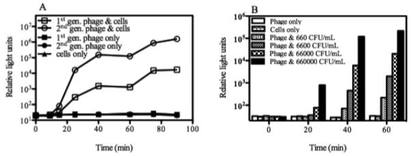
Fig. 1.
Reporter phage-mediated bioluminescent detection of Y. pestis. A. Signal response time and signal strength. The ability of the first and second generation (1st gen., and 2nd gen.) reporter phages to transduce a bioluminescent signal response to Y. pestis was compared. Y. pestis was grown in Luria Bertani broth at 28°C to an A600 of approximately 0.2. At time 0, cells (~1 × 10⁸ CFU/mL) were mixed with the reporter phage (multiplicity of infection [MOI] of 0.5) and incubated at 28°C. Bioluminescence (RLU) was measured over time following the addition of the substrate n-decanal. Numbers are the mean (n=3) ± SD. B. Dose-dependent detection. 10-fold serial dilutions of a Y. pestis culture were mixed with the second generation reporter phage and incubated at 28°C. Bioluminescence (RLU) was measured over time following the addition of n-decanal. Numbers are the mean (n=3) ± SD.

