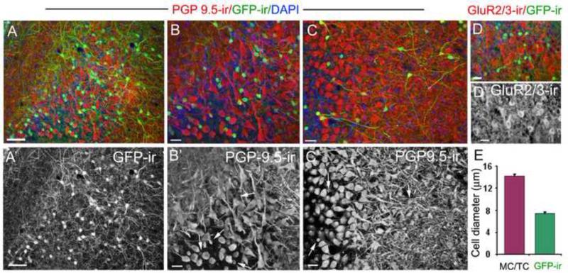
Figure 7. The cholinergic (GFP-ir) interneurons in the mitral cell layer are not mitral/tufted cells.
A: A representative image of GFP-ir and PGP 9.5 immunoreactivity (PGP9.5-ir) in the EPL and MCL. The PGP 9.5 antibody strongly labeled mitral/tufted cells and their nerve processes (red), as well as GFP-ir cell bodies. A’: Image of GFP-ir alone. B and B’: An enlarged image showing the MCL region in panel A (lower middle portion) and PGP9.5-ir alone, respectively. The GFP-ir signal (green) in panel B is reduced using Adobe Photoshop for better visualization of the cell bodies. The GFP-ir cells exhibit smaller cell bodies and thin nerve processes, whereas mitral cells are multi-polar with large cell bodies and thick nerve processes. None of the mitral cells are positive for GFP-ir. C and C’: Cholinergic interneurons (GFP-ir) and PGP9.5-labeled mitral/tufted cells viewed from a different angle. GFP-ir cell bodies in the deep EPL and the MCL intermingle with mitral/tufted cell bodies and processes. Some GFP-ir cells show long nerve processes. Representative GFP-ir cell bodies are pointed by arrows in B’ and C’. D: A representative confocal image of GluR2/3 immunoreactivity (GluR2/3-ir) in mitral/tufted cells and GFP-ir. D’: GluR2/3-ir alone. Stronger GluR2/3-ir is seen in cell bodies as compared to the surrounding nerve processes. There is no colocalization between GFP-ir and GluR2/3-ir. DAPI (blue) in A, B, C and D. E: Average cell diameters measured from GFP-ir cells and cells labeled with PGP 9.5-ir or GluR2/3-ir. For mitral/tufted cells, only those with recognizable morphological features were measured (22 cells for each group, respectively). Scale: A, 50μm. B to E, 20μm.

