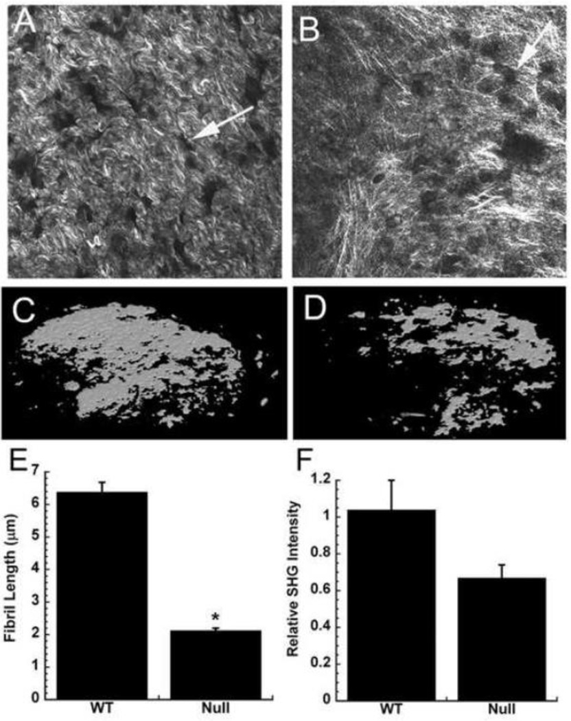
Figure 1. Osteonectin-null bone matrices synthesized in vitro are disorganized and hypomineralized.
Collagen fibrer morphology in wild type (A) and osteonectin-null (B) matrices, imaged by SHG (representative fields, individual optical sections). Arrows indicate sites in the matrix formerly occupied by osteoblasts. MicroCT imaging of mineral in wild type (C) and osteonectin-null (D) matrices (representative image of entire well). Collagen fiber length (E) and relative SHG intensity (F) in wild type vs. osteonectin-null matrix. * = significantly different from WT, p<0.01.

