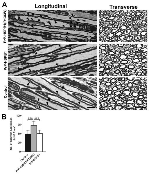
Figure 3.
One μm-thick, toluidine blue stained longitudinal and cross sections from mid-sciatic nerves of 1 year old PrP-HSPB1(R136W), PrP-HSPB1 and non-transgenic controls. (A) SLI is marked by arrows on the longitudinal sections; ‘S’ donates axons with SLI on the cross sections. Numerous SLIs are present in PrP-HSPB1(R136W). Bar = 10 μm for all longitudinal and 10 μm for all cross sections. Density of SLI on the cross sections of sciatic nerves from PrP-HSPB1(R136W), PrP-HSPB1 and non-transgenic controls is shown in panel B. The number of SLI determined from a total area of 0.712 mm2 is significantly increased in the PrP-HSPB1(R136W) mice compared to PrP-HSPB1 and non-transgenic controls (***p <0.001).

