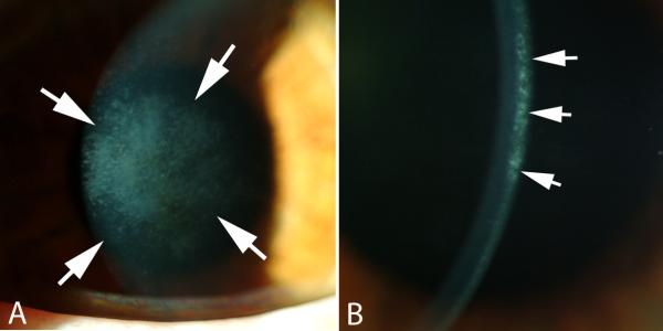
Fig. 1.
Corneal stromal opacity (haze). A shows haze that overlies the area of ablation with the excimer laser that occurred at three months after PRK for -7-diopters of myopia when no mitomycin C treatment was applied at the time of surgery. Magnification 20X. B. Slit lamp examination shows that the haze is localized in the anterior stroma immediately beneath the epithelial basement membrane. Magnification 40X.

