Fig. 3.
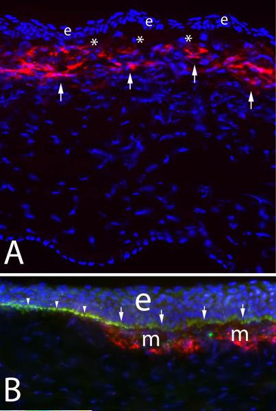
Corneal myofibroblasts. A. Myofibroblasts (arrows) beneath the basement membrane in the anterior stroma after PRK for -9 diopters of myopia in a rabbit cornea. e indicates epithelium. * is artifactual breaks between the epithelium and stroma that occur during tissue sectioning. 200X magnification. B. At higher magnification with double staining for the α-smooth muscle actin maker for myofibroblasts (red) and integrin beta-4 (green) decorating the epithelial basement membrane in a rabbit cornea that had -9 diopter PRK, it can be seen that the basement membrane appears more irregular (arrows) overlying the myofibroblasts (m) than it does in the adjacent area free of myofibroblasts (arrowheads). e indicates the epithelium. The blue is DAPI staining for all cell nuclei. Magnification 600X. B reprinted by permission from Netto MV, Mohan RR, Sinha S, Sharma A, Dupps W, Wilson SE. Stromal haze, myofibroblasts, and surface irregularity after PRK. Exp. Eye Res. 2006;82:788-97.

