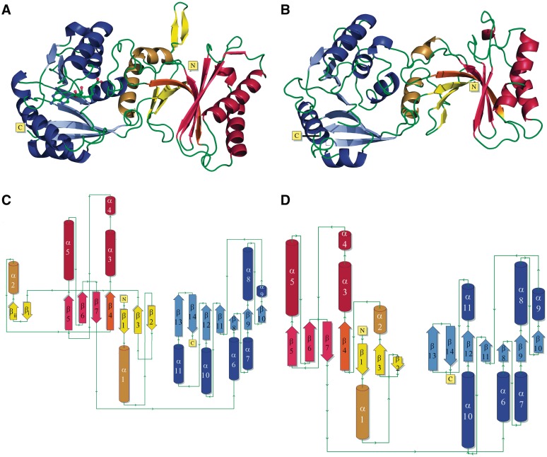
Figure 2.
Crystal structures of PfTrm14 and TTCTrmN. (A and C) show the structure of PfTrm14 in cartoon representation (A) and as a topology diagram (C); (B and D) show the structure of TTCTrmN in cartoon representation (B) and as a topology diagram (D). β-strands are shown in red, yellow and blue for the core-THUMP, NFLD and RFM (sub)domains, respectively. α-Helices are shown in dark red, yellow and blue. The β-strand shared by the core-THUMP and NFLD subdomains is shown in orange. The two additional β-strands in the PfTrm14 structure are named βi and βii. The structure of PfTrm14 contains sinefungin bound in the active site, represented in ball and stick.

