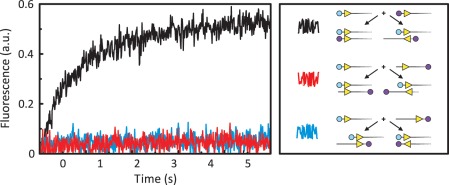
Figure 2.
FRET from synapsis in trans. Equal volumes of FokI endonuclease and a solution containing two DNA molecules were mixed in the stopped-flow fluorimeter to give reactions in C-buffer at 20°C with FokI at 25 nM and both DNA at 12.5 nM. Excitation was at 545 nm and the change in emission (in arbitrary units: a.u.) at >645 nm recorded over time. The black, red and blue traces each came from a reaction with a particular pair of DNA molecules, as indicated in the right-hand panel. The DNA molecules were 260 bp long and contained a single recognition site for FokI (yellow arrowhead) 30 bp from the nearest end of the DNA: the arrowheads mark the orientation of the recognition sequence by pointing towards the downstream sites for DNA cleavage. For each experiment, one of the DNA molecules was labelled at one end with Ax546 (cyan circles) whereas the other DNA was labelled with Ax647 (mauve circles) at either the same or the opposite end. The pairs of the substrates that gave rise to the black, the red and the blue trace are depicted next to the squiggle of the same colour. The schemes below each pair show the structures formed by parallel and anti-parallel synapses of the FokI sites on both substrates within that pair: the relative locations of the Ax546 and Ax647 dyes are indicated.

