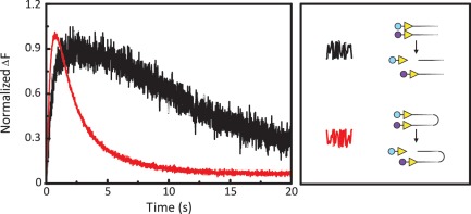
Figure 4.
FRET from cutting one-site and two-site DNA. Equal volumes of FokI endonuclease and DNA labelled with Alexa Fluor dye(s) were mixed by stopped-flow to give the following reactions in M-buffer at 20°C. One reaction (black trace), contained 25 nM FokI and two DNA molecules with one FokI site, both at 12.5 nM: the DNA was labelled at the end proximal to the FokI site, one with Ax546 and the other with Ax647. The second reaction (red trace) contained FokI (50 nM) and one DNA molecule (25 nM) with two inverted FokI sites, labelled at one end with Ax546 and at the other with Ax647. [All DNA were 260 bp long, with 30 bp from the FokI site(s) to the proximal end(s). The locations and orientations of the recognition sites (yellow arrowheads) and the fluorescent labels (Ax546, in cyan; Ax647 in mauve) on the substrates are indicated in the right-hand panel, as are also representative cleavage products.] Excitation was at 545 nm and the change in emission at >645 nm recorded over time. Since the emission from the reaction on the one-site substrates was lower than that from the two-site substrate, the fluorescence signal (ΔF) is shown normalized to a value of 1 for the maximal signal from each reaction.

