Figure 1.
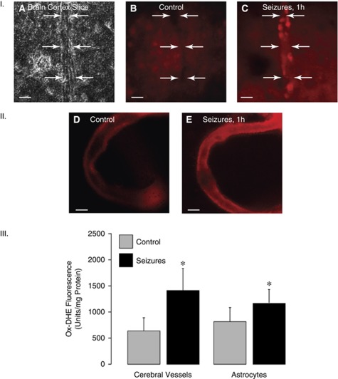
Effects of seizures (1 hour) on reactive oxygen species (ROS) in cerebral vessels and cortical astrocytes as detected by dihydroethidium (DHE) oxidation (ox-DHE). Panel I: Ox-DHE fluoromicrography of the brain cortex slices. (A) A phase contrast image of a brain cortex slice with a penetrating vessel; (B) a fluorescence image, control brain; (C) a fluorescence image, seizing brain (representative pictures; bar, 200 μm). Arrows indicate the outer borders of the vessel wall. Panel II: Ox-DHE fluoromicrography of pial arterioles excised from control (D) and seizing (E) piglets 1 hour after saline (D) or bicuculline (E) injection (representative pictures; bar, 200 μm). Panel III: Detection of ox-DHE by fluorescence spectroscopy of cortical cerebral microvessels (60 to 300 μm) and cortical astrocytes (N=8 animals in each group). *P<0.05 compared with control values.

