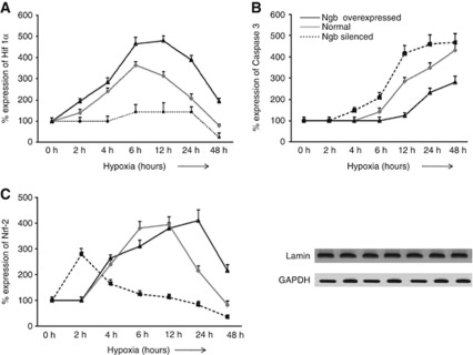
Figure 7.
Neuroglobin (Ngb) overexpression delays apoptosis in N2a cells exposed to hypoxia. Ngb-silenced, Ngb overexpressed, and normal N2a cells were exposed to different durations of hypoxia. Western blots were performed with nuclear fractions to study the expression of hypoxia-inducible factor-1α (Hif-1α) and nuclear factor erythroid 2-related factor 2 (Nrf2) while the cytosolic fraction was used to study caspase 3 expression. Protein expression was calculated in percentage, considering optical density of protein bands at 0 hour hypoxia to be 100%. Points on the graph depict mean±s.e.m. of six individual western blot observations at a particular duration of exposure to hypoxia. Graphs depict percentage protein expression of (A) Hif-1α, (B) Nrf2, and (C) caspase 3 following different durations of exposure to hypoxia. Lamin and glyceraldehyde phosphate dehydrogenase (GAPDH) were used as loading controls for nuclear and cytosolic proteins, respectively.

