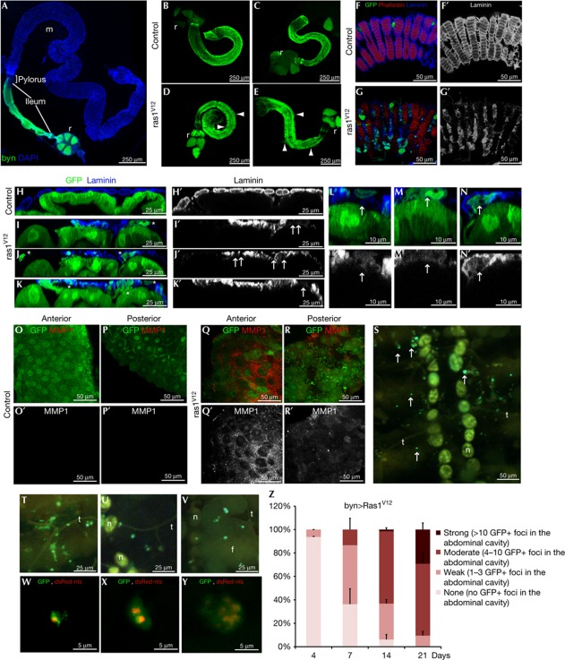
Figure 1.
Ras1V12 expression is sufficient to initiate dissemination of the adult Drosophila hindgut cells. (A) Drosophila intestinal epithelium. Hindgut cells green fluorescent protein (GFP)-marked using byn-GAL4. Blue, nuclei (DAPI); m, midgut, r, rectum. (B–E) Hindguts expressing GFP alone (B,C) or GFP plus Ras1V12 (D,E) 7 days (B,D) or 21 days (C,E) after induction of transgenes. Arrowheads: cellular protrusions throughout ileum. (F,G) Surface views of ileum expressing GFP alone (F) or GFP plus Ras1V12 (G). Blue, laminin (basement membrane). (F′,G′) Laminin channel only of F and G. (H–K′) Cross-sections of control (H,H′) and Ras1V12 (I–K′) hindguts. Ras1V12-expressing hindguts (I–K) show GFP+ cells in the process of invading basally out of the gut epithelium (stars). These regions also have reduced or absent laminin staining (I′–K′, arrows). (L–N) Close-up views of disseminating cells (arrows) from Ras1V12-expressing hindguts. (laminin, blue). (L′,N′) Laminin channel only of L–N. Disseminating cells are present in regions of reduced laminin staining (arrows). (O–R) Matrix metalloprotease 1 (MMP1) expression (red) in control (O,P) and Ras1V12-expressing hindguts (Q,R). (O′–R′) MMP channel only of O–R. (S) Inside view of the abdominal cavity from an animal expressing Ras1V12 in the hindgut showing GFP+ foci (arrows). Stereoscope (T–Y) and confocal (W–Y) live views of GFP+ foci and dsRed nuclei (dsRed-nls). Note: yellow autoflorescence makes nephrocytes (n), fat body (f) and trachea (t) visible. (Z) Quantification of Ras1V12-induced dissemination over time. DAPI, 4,6-diamidino-2-phenylindole; nls, nuclear localization signal.

