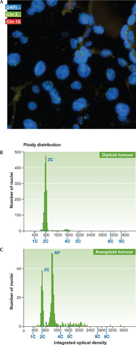
Figure 3.
Direct methods to assess tumour CIN status. (A) Section from a renal cell carcinoma biopsy, hybridized to two fluorescently labelled centromere probes—chromosome 2 (green) and chromosome 15 (red). Variation in centromere copy number is evident between the nuclei. (B) DNA image cytometry profile for a diploid tumour (diploid DNA content determined relative to control diploid cells). (C) DNA image cytometry profile for an aneuploid tumour (determined relative to control diploid cells).

