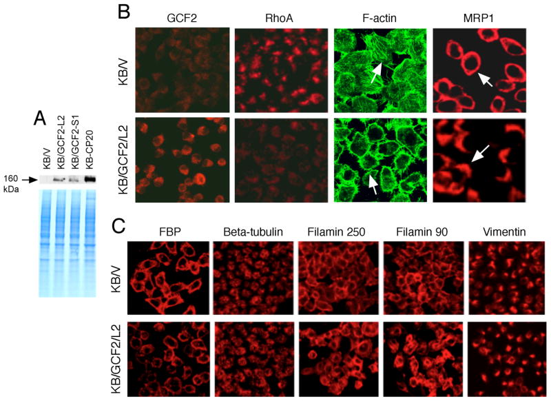
FIGURE 3.
Overexpression of GCF2 in GCF2-transfected cells. (A) Immunoblot showing increased expression of GCF2 in two clones of GCF2-transfected cells, KB/GCF2/L2 and KB/GCF2/S1, compared with KB/V cells that were transfected with vector only. The cell line most resistant to cisplatin, KB-CP20, served as a positive control. The lower panel is a Coomassie blue-stained gel, serving as a loading control. (B) Confocal immunofluorescence analysis of expression, distribution, and localization of GCF2, RhoA, F-actin, and MRP1. KB/V was transfected with vector only, serving as controls; KB/GCF2/L2 is a clone of GCF2-transfected cells. (C) Confocal images reveal differences between KB/V and KB/GCF2/L2 cells in expression, distribution, and organization of FBP, β-tubulin, filamin 250, filamin 90, and vimentin, in KB/V and KB/GCF2/L2, as described above.

