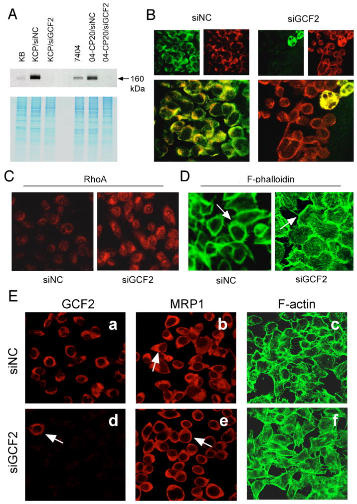
FIGURE 6.
siRNA of GCF2 reverses phenotypes of CP-r cells. (A) Immunoblot revealing suppression of GCF2 expression in siGCF2-transfected cells, labeled as KB-CP20/siGCF2, and 04-CP20/siGCF2 cells, in comparison to their negative control siNC, labeled as KB-CP20/siNC, and 7404-CP20/siNC, respectively. (B-D): KB-CP20 cells, (B) Double-stained confocal images (larger, lower panels) show that siGCF2 silenced the expression of GCF2 (stained with green, upper left), and MRP1 (stained with red, upper right) re-appeared at the cell surface as seen at the right, and two giant multinucleated cells at the top right show a mixed pattern of double-fluorescence, and seemed to not be affected by the siGCF2. siNC represents siRNA of a negative control (mock-transfected cells), seen at the left. (C) Expression of RhoA was recovered in the siGCF2-transfected cells (on the right, labeled as siGCF2) after GCF2 was knocked-down by the specific siRNA of GCF2, in comparison to the mock cells seen on the left, labeled siNC. (D) F-phalloidin stained immunofluorescence images indicate fine fibers were re-formed on the cell surface of siGCF2-transfected cells compared to siNC. (E) Human liver carcinoma cisplatin-resistant 7404-CP20 cells were transfected with siGCF2, labeled as siGCF2, or with siRNA as a negative control, labeled as siNC. In the GCF2 panels, an arrow points to the single cell in this field still expressing GCF2, while all other cells show no signal, as expression of GCF2 was inhibited by the siRNA of GCF2. In the MRP1 panels, an arrow points to cap-like staining of MRP1 in siNC-transfected cells (upper panel), and an arrow indicates MRP1 was re-located to the cell surface (lower panel). In the F-actin panels, the fine filaments were visualized at the cell surface in siGCF2-transfected cells (lower panel) compared to siNC mock-transfected cells in which the cells appear as hollow circles (upper panel).

