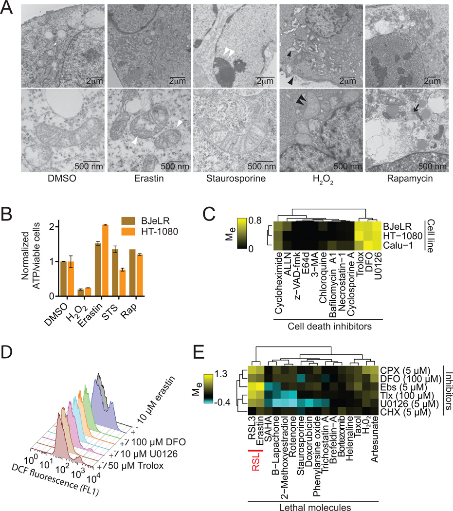
Figure 2. Erastin-induced oxidative death is iron-dependent.
(A) Transmission electron microscopy of BJeLR cells treated with DMSO (10 hrs), erastin (37 µM, 10 hrs), staurosporine (STS, 0.75 µM, 8 hrs), H2O2 (16 mM, 1 hr) and rapamycin (Rap, 100 nM, 24 hr). Single white arrowheads: shrunken mitochondria; paired white arrowheads: chromatin condensation; black arrowheads: cytoplasmic and organelle swelling, plasma membrane rupture; black arrow: formation of double-membrane vesicles. A minimum of 10 cells per treatment condition were examined. (B) Normalized ATP levels in HT-1080 and BJeLR cells treated as in (A) with the indicated compounds. Representative data (mean+/−SD) from one of three independent experiments is shown. (C) Modulatory profiling of known small molecule cell death inhibitors in HT-1080, BJ-eLR and Calu-1 cells treated with erastin (10 µM, 24 hrs). (D) Effect of inhibitors on H2DCFDA-sensitive ROS production in HT-1080 cells treated for 4 hours. (E) Modulatory profiling of ciclopirox olamine (CPX), DFO, ebselen (Ebs), trolox (Tlx), U0126 and CHX on oxidative and non-oxidative lethal agents.

