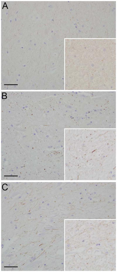
Fig 3.

Photomicrograph. (a) Control animal; iNOS immunoreactivity is faint and restricted to microglial processes (inset); (b) Experimental animal; iNOS immunoreactivity is increased and punctate with large globular regions of immunoreactivity in microglial processes at the site of the cervical trauma (inset); (c) Experimental animal; iNOS immunoreactivity is increased in microglial processes in the lumbar spinal cord section (inset). Hematoxylin and eosin, bar = 0.5mm.
