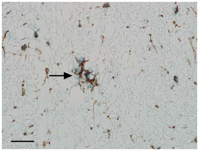
Fig 4.

Photomicrograph. Representative image of the affected spinal cord illustrating that microglia are stained with IBA-1 (brown) and iNOS (blue) indicating dual expression of these markers. Immunoperoxidase staining, DAB chromagen (IBA-1) and Vector blue (iNOS), Mayer’s hematoxylin counterstain, bar = 100μm.
