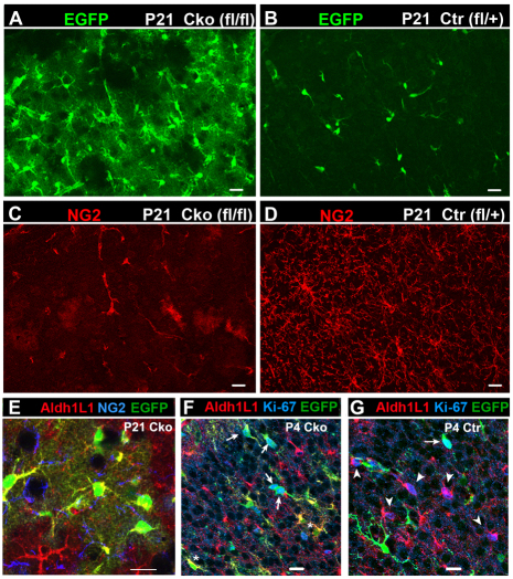
Fig. 2.
NG2 cell-derived astrocytes in the dorsal forebrain of P21 Olig2 Cko and Ctr mice. (A-D) EGFP fluorescence (green, A,B) and NG2 immunolabeling (red, C and D) of the neocortex of P21 Olig2 Cko (A,C) and Ctr (B,D) mice. Upper left is pial surface. NG2+ EGFP+ polydendrocytes in the Ctr cortex are completely replaced by NG2-negative EGFP+ protoplasmic astrocytes in Cko brain, where NG2 immunoreactivity is detected only in the vasculature (C). (E) Coronal section through P21 Cko neocortex stained for Aldh1L1 (red) and NG2 (blue). EGFP+ NG2 cell-derived protoplasmic astrocytes are Aldh1L1+. (F,G) EGFP fluorescence (green) and immunolabeling for Aldh1L1 (red) and Ki-67 (blue) in the neocortex of Cko (F) and Ctr (G) mice at P4. In Cko neocortex, Ki-67 is detected mostly in EGFP+ cells (arrows in F) but rarely in EGFP– Aldh1L1+ cells, and some EGFP+ cells express Aldh1L1 (asterisk in F). In Ctr mice, many EGFP– Aldh1L1+ resident astrocytes are Ki-67+ (arrowheads in G). Occasional EGFP+ Ki-67+ cells are seen in Ctr (arrow in G). Scale bars: 20 μm.

