Fig. 1.
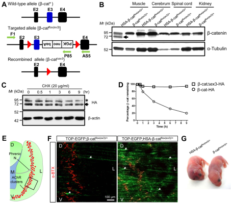
Increased stability and activity of β-catenin in skeletal muscles in HSA-β-catflox(ex3)/+ mice. (A) Schematic of the wild-type and targeted β-catenin allele. E, exon; red triangle, loxP sequence; green arrows, primers for genotyping. (B) Specific expression of exon 3-deleted β-catenin (arrow) in skeletal muscles of HSA-β-catflox(ex3)/+ mice, but not in other tissues. (C) Increased stability of exon 3-deleted β-catenin compared with wild type. HEK293 cells were transfected with HA-tagged wild-type (asterisk) and mutant (arrow) β-catenin, and treated with 20 μg/ml CHX for various times. β-Actin was used as a loading control. (D) Quantification of data in C (mean ± SEM, n=3). (E) Schematic of the left hemi-diaphragm. Red dots, AChR clusters; rectangle, area shown in F; D, dorsal; V, ventral; L, lateral; M, medial. (F) Expression of TOP-EGFP in diaphragm muscles by β-catenin GOF mutant. Muscles of control (TOP-EGFP;β-catflox(ex3)/+) and TOP-EGFP;HSA-β-catflox(ex3)/+ mice were stained with Alexa Fluor 594-conjugated α-BTX (red). Image was taken by confocal fluorescence microscope. Area shown is indicated by rectangle in E. (G) Neonatal mice of indicated genotypes.

