Fig. 7.
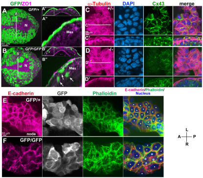
Loss of Sox17 results in defective epithelial polarity and disorganized cellular adhesion. (A-B″) GFP and ZO1 IHC in Sox17GFP/+ and Sox17GFP/GFP mouse embryos at 3 ss (A,B). Optical transverse sections from the reconstructed confocal images showed a compact epithelial layer of definitive endoderm cells in Sox17GFP/+ embryos (A′,A″), whereas in Sox17GFP/GFP embryos the epithelial layer was uneven and a portion of the GFP-positive cell population was found interspersed among the mesoderm cells (B′,B″). (C-D′) Co-IHC of α-tubulin and Cx43 together with nuclear staining in Sox17+/– and Sox17–/– embryos at 3 ss. Microtubules labeled with α-tubulin showed a parallel arrangement in Sox17–/– endoderm (star, D), in contrast to the radial arrangement in Sox17+/– embryos, accompanied by a reduction in the membrane localization of Cx43 (optical transverse sections in C′,D′). (E,F) E-cadherin, GFP and actin distribution in endoderm near the node. Note the disorganized localization of E-cadherin in the Sox17GFP/GFP embryos (F) as compared with the control embryos (E), suggesting weak adherens junctions. Stars indicate individual cells; the blue stars indicate cells with much reduced E-cadherin distribution in the cell boundary. Mes, mesoderm; N, node.

