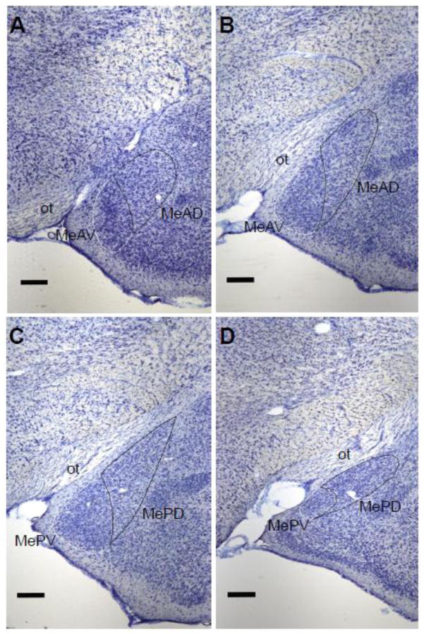
Figure 4.
Boundaries of the Me quadrants as determined in Nissl-stained coronal sections. A: The most rostral section containing MeAD and MeAV as defined by the differential orientation of cells extending dorsomedially. B: The most caudal section containing MeAD and MeAV as defined by the optic tract (ot) extending midway to the dorsal tip of the MeAD, while the MeAD still maintains a round shape. C: The most rostral section containing MePD and MePV as defined by the ot extending to the dorsal tip of the MePD, while the MePD forms a point at the most dorsal end. D: The most caudal section containing both MePD and MePV defined as the MePD starting to round out and move more dorsomedial, while the MePV remains a distinct small round group of cells. Scale bar = 250 μM.

